

Active pharmaceutical ingredients (API)
From in vitro ADME/DMPK assays to physiologically based pharmacokinetic modelling, Eurosafe secures the development of your drug candidates and supports the pharmaceutical, biotech, chemical and agrochemical industries.
In vitro assays
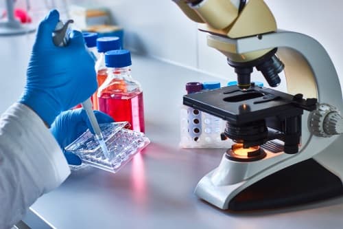
Our in vitro ADME and DMPK assays relate to solubility, in vitro metabolism, in vitro permeability and transporters, binding to proteins and bioanalysis.
Our ADME services comply with external guidelines (GLP, FDA, EMA, OECD) or are performed in a screening mode.
Eurosafe serves a number of industries including big pharma, biotech, as well as chemical and agrochemical industries.
Metabolism
Metabolite profiling and identification using high resolution mass spectrometry, and eventually quantification.
It is well known that the metabolism of a given test compound often contributes to its efficacy and toxicity observed in vivo.
Assays are standardized for:
- metabolite identification,
- metabolite profiling,
- metabolite stability,
- identification of metabolizing enzymes,
- stability in plasma and buffer (part of BCS evaluation),
- PBPK modeling.
In vitro metabolism studies conducted in biological matrices ranging from isolated cells to subcellular fractions allow the evaluation of intrinsic metabolic potential or specific mechanisms with respect to a given reaction.
Eurosafe recommends the incubation of liver, intestine and skin microsomes, as well as hepatocytes, HepaRG™, expressed enzymes and plasma.
In addition, in vitro systems derived from various animal species permit the comparison of metabolic pathways among species (including humans) before a given compound can be tested clinically. Metabolites from Phase I and Phase II enzymes are scanned.
DDI — Induction
Cytochrome P450 induction forms part of in vitro experimental ADME services. A new test compound's induction profile should routinely be evaluated.
These studies include but are not limited to:
- CYP1A2, CYP2B6 and CYP3A4,
- other Phase I enzymes including monoamine oxidase (MAO), flavin monooxygenase (FMO), xanthine oxidase (XO), and alcohol/aldehyde dehydrogenase,
- Phase II enzymes including UDP glucuronosyl transferases (UGTs).
Human hepatocytes continue to serve as the system of choice for evaluating enzyme induction in vitro, both with freshly isolated human hepatocytes and cryopreserved hepatocytes available for routine use.
When determining the enzyme induction potential of new test compounds using cultured human hepatocytes or HepaRG™ cells, the following elements must be considered:
- inter-individual variability,
- changes in mRNA level of target genes as an endpoint (activity level is also evaluated at Eurosafe),
- the inclusion of vehicle controls, positive controls (usually known strong inducers), and negative controls (usually known non-inducers).
Initially CYP1A2, CYP2B6, and CYP3A induction is evaluated. If no induction of CYP3A4/5 enzymes is observed, it is unnecessary to evaluate the induction potential of CYP2C enzymes because both are induced via activation of the pregnane X nuclear receptor (PXR). If CYP3A4/5 induction does occur, it is then necessary to evaluate the potential of CYP2C induction.
We examine fold-change in CYP enzyme mRNA levels when incubated with the test compound by using the cutoffs described in the FDA guidelines.
DDI — Inhibition
Inhibition of metabolism enzymes (CYP and UGT) by a new test compound may decrease the metabolism of co-medicated drugs. The potential of this test compound to inhibit enzymes is usually investigated to elucidate the precise inhibition mechanisms (reversible or time-dependent) as well as inhibition potency (e.g. Ki).
Liver microsomes are recommended for screenings and for mechanistic evaluations. Alternatively, human hepatocytes or HepaRG™ cells can be used to determine the CYP inhibition potential of test compounds in the cellular environment.
Screening assays are performed with up to 9 cytochrome P450 enzymes (1A2, 2A6, 2B6, 2C8, 2C9, 2C19, 2D6, 2E1, 3A4/5) using 10 probe reactions. The direct inhibition assay delivers IC50 values towards each CYP enzyme; the time-dependent inhibition assay yields direct and shifted IC50 values.
Following IC50 determination, we determine Ki for the test compound against the appropriate enzyme. This parameter elucidates the potency of inhibition as well as its type (competitive, non-competitive, uncompetitive or mixed), and can be used to estimate the impact of any potential in vivo interaction.
DDI — Phenotyping
In order to predict DDI, it is crucial to understand which enzymes (Phase I and Phase II) are responsible for the metabolism of a given test compound.
There are three well-characterized methods recognized by the FDA for identifying the individual CYP enzymes involved in a drug's metabolism:
- method 1 uses a bank of human liver microsomes characterized for CYP activity,
- method 2 uses individual human recombinant CYP enzymes,
- method 3 uses specific chemicals or antibodies as specific enzyme inhibitors,
- method 3 bis: Eurosafe proposes the use of Silensomes™.
Silensomes™ are validated human-pooled liver microsomes (HLMs) which are chemically and irreversibly inactivated for a specific CYP450 using mechanism-based inhibitors (MBI). This approach allows quantitative phenotyping by determining metabolic contribution (fm) instead of RAF evaluation.
Y. Parmentier, C. Pothier, A. Delmas, F. Caradec, M.-M. Trancart, F. Guillet, B. Bouaita, C. Chesné, J. B. Houston & B. Walther, “Direct and quantitative evaluation of the human CYP3A4 contribution (fm) to drug clearance using the in vitro SILENSOMES model”, Xenobiotica, vol. 47, pp. 562-575, 2016.
Transporters
Evaluating transporter-mediated drug interactions.
Membrane transporters have clinically relevant effects on the pharmacokinetics and pharmacodynamics of drugs in various organs and tissues by controlling the absorption, distribution, and elimination of drugs (Giacomini, Huang et al., 2010; Giacomini and Huang, 2013).
Several transporters interact with clinically relevant drugs — see the FDA guidelines. For example:
- P-glycoprotein (P-gp or Multi-drug Resistance 1, MDR1),
- breast cancer resistance protein (BCRP),
- organic anion transporting polypeptide 1B1/1B3 (OATP1B1/OATP1B3),
- organic anion transporter 1/3 (OAT1/OAT3),
- multidrug and toxin extrusion (MATE) proteins,
- organic cation transporter 2 (OCT2).
Understanding whether a certain drug behaves as a substrate or perpetrator (inhibitor or inducer) of these key transporters helps explain clinical consequences (toxicity or altered efficacy) resulting from altered tissue distribution.
Coupled with appropriate in vitro-to-in vivo extrapolation methods such as PBPK, these assays help determine whether the sponsor should conduct in vivo drug interaction studies.
Permeability (Caco-2)
Permeability assay for predicting the in vivo absorption of drugs across the gut wall by measuring the rate of transport of a compound across the Caco-2 cell line.
The Caco-2 permeability assay is part of our portfolio of in vitro ADME screening services. To understand the suitability of a test compound for oral dosing and to predict the absorption of orally administered drugs, the Caco-2 assay is widely used across the pharmaceutical industry as an in vitro model of the human small intestinal mucosa.
The Caco-2 cell model mimics processes such as transcellular transport, paracellular transport, and some aspects of efflux and active transport. Assessing transport in both directions (apical to basolateral and basolateral to apical) across the cell monolayer yields an efflux ratio.
Monolayers based on Caco-2 cells which express MDR1 and BCRP transporters allow the study of passive permeability, if the test compound functions as substrate or inhibitor of those transporters.
Based on in vitro permeability and solubility data, the Biopharmaceutics Classification System (BCS) is a regulatory mechanism through which one can obtain a waiver of clinical bioequivalence studies, also called a biowaiver. According to the FDA BCS guidance, compounds classified as Class I (highly soluble and highly permeable) are eligible for BCS biowaivers.
Protein binding
Identification of the free fraction of drugs on plasma proteins, cells, or sub-cellular fractions.
Eurosafe offers different approaches to evaluate plasma protein binding:
- equilibrium dialysis method: the most widely used method for assessing drug binding, where non-specific fixation is kept at a minimum;
- sharing between erythrocytes and plasma proteins: applied when the test compound is lipophilic or the non-specific binding is high.
Studying a drug's binding to different plasma proteins and erythrocytes also helps evaluate its blood distribution. This distribution helps elucidate a drug's binding capacities (NKa), and hence enables the simulation of human pathophysiological conditions.
Eurosafe performs ex vivo studies to correlate the plasma binding percentage of drug candidates (with high affinities for HDL) in 50 plasma samples derived from a pharmacokinetic study with the concentration of a given lipoprotein (p < 0.0001).
Eurosafe also evaluates drug binding to microsomal proteins and cells in order to evaluate the free fraction (fumic, fuhep) and to predict hepatic clearance or drug-drug interaction potential in vivo, using in vitro microsomal metabolic data.
Transcutaneous passage
Evaluation of the dermal absorption and delivery of a test substance using excised skin.
In vitro methods measure the diffusion of chemicals across skin into a fluid reservoir. Cryopreserved skin is used to measure diffusion only, whereas fresh metabolically-active skin is used to simultaneously measure diffusion and skin metabolism.
Such methods have found particular usage as screening tests for comparing transcutaneous delivery of chemicals from different formulations. Acceptable data from a minimum of four replicates per test preparation are required.
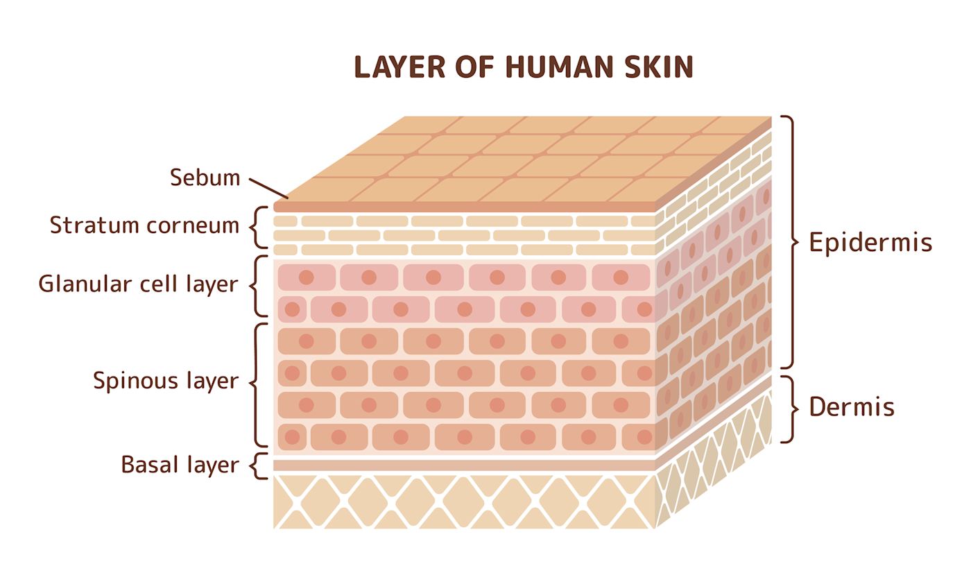
Using the conditions described in the OECD 428 guidelines, absorption of test compounds during a given time interval is measured by analyzing both receptor fluid and the treated skin. All components of the skin should be analyzed and recovery determined; in some cases, the skin may be fractionated into stratum corneum, epidermis, and dermis fractions for additional analysis.
In silico studies / Computational toxicology
Our in silico services expedite the prediction of biological properties related to toxicity, activity and ADME, as well as high-throughput screening and lead identification.
We also perform physiologically based pharmacokinetic (PBPK) modelling on state-of-the-art computing facilities.
Structure modeling
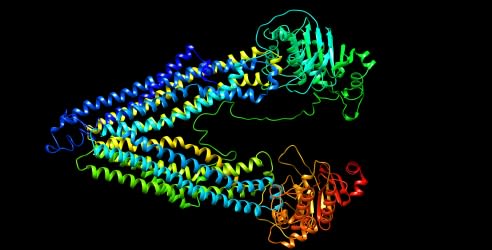
Eurosafe offers services for the prediction of the three-dimensional structure of proteins of interest by the homology modeling method, using automated state-of-the-art methods. Homology modeling uses the sequence similarity of amino acid residues with well-known 3D structures (by X-ray and NMR) to predict the structure of an unknown protein.
The structural models are validated for quality and further optimized by molecular dynamics simulation. The optimized model can be used for molecular docking, macromolecular engineering and drug design.
Homology modeling service includes:
- sequence alignment of amino acids,
- prediction of the 3D model,
- structure validation,
- structure optimization by molecular dynamics.
Molecular docking
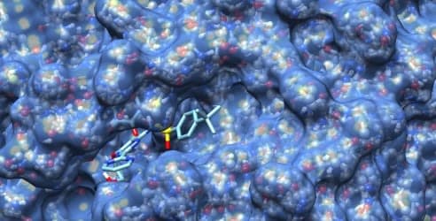
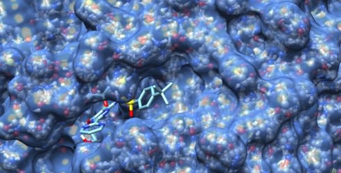
Eurosafe offers a molecular docking service to discover the interaction between the protein of interest and a ligand by an automated high-throughput screening algorithm. We report binding affinity, ligand conformation search and the active site of the protein. On demand, the protein-ligand complex is further optimized by molecular dynamics simulation for interaction refinement.
Molecular docking service includes:
- structure preparation of protein and ligand,
- automated docking simulation,
- affinity prediction, ligand pose prediction and active site prediction,
- protein-ligand complex optimization by molecular dynamics.
QSAR
The Eurosafe QSAR service includes the prediction of relationships between the geometric structure of a chemical compound and its biological activity. We use QSAR methods such as the OECD Toolbox, VEGA and Toxtree to predict the skin sensitization potential of compounds, according to OECD guidelines (ENV/JM/MONO(2007)2).
QSAR service includes:
- biological properties prediction,
- skin sensitization prediction of ingredients,
- skin metabolism and sensitization prediction of ingredients,
- safety assessment of compounds.
PBPK
Eurosafe offers a physiologically based pharmacokinetic (PBPK) modeling service to predict the pharmacokinetics of compounds, using the validated open-source software PK-Sim.
We offer PBPK services for the interpolation and extrapolation of knowledge from in vitro pharmacological or toxicological experiments to in vivo, between:
- effect of dose and exposure duration: e.g. from continuous to discontinuous, or single to multiple exposures,
- effect of routes of administration: e.g. from inhalation exposures to ingestion,
- species and individual variation in the pharmacokinetics of compounds,
- in vitro to in vivo extrapolation (IVIVE): the qualitative or quantitative transposition of experimental results to predict phenomena in biological organisms,
- study of drug-drug interactions (DDI),
- study of the ADME compartmental model predictor of absorption, distribution, metabolism and excretion.